A clearer picture of the heart: Imaging that puts patients first
A University of Ottawa-based innovation advanced cardiac PET scans, benefiting patients in Canada and around the world.

At age 13, Everad Tilokee was diagnosed with dilated cardiomyopathy, a condition that weakens the heart and restricts blood flow. Motivated to learn more about this condition, Everad later pursued a master’s degree focused on cardiac stem cells at the University of Ottawa Heart Institute.
In 2014, Everad received a life-saving heart transplant and returned to the Institute—this time as a patient. Heart transplant recipients receive lifelong monitoring for signs of coronary disease and transplant related complications. For some, the standard cardiac test is an angiogram, a diagnostic procedure using X-Ray and a catheter inserted through the wrist or thigh to show how the blood flows through the heart.
At the University of Ottawa Heart Institute, Everad instead received a cardiac positron emission tomography (PET) perfusion. This procedure creates images of the heart’s blood vessels using a scanning machine and an injection of a low dose of radioactive isotope called rubidium‑82.
Both procedures are effective, but Everad would feel sore for days after doing an angiogram. “With PET imaging, it’s just an IV—no pain, no recovery time,” he says. “It’s a night and day difference in how I live my life.”
Today, this less invasive procedure is a standard used to monitor hundreds of thousands of at-risk patients worldwide. But few people know that this clinical standard of care emerged from CIHR-funded research developed right here in Canada, at the University of Ottawa Heart Institute.
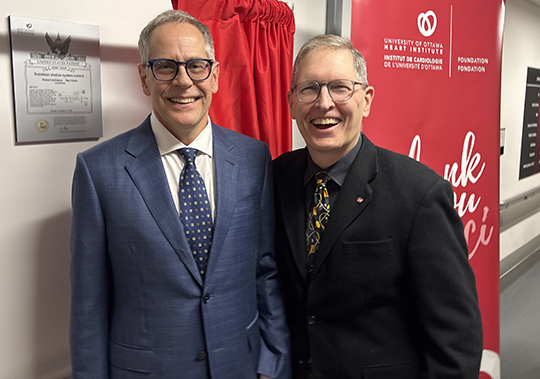
Dr. Robert deKemp, a physicist specialized in cardiac imaging, and Dr. Robert Beanlands, a clinician-scientist at the time (now President and CEO of the University of Ottawa Heart Institute) joined forces in 2009 to help address a pressing global challenge: the shortage of technetium-99m, a critical isotope used in nuclear medicine such as imaging of the heart, lungs, kidneys, and tumours.
Repeated and prolonged closure of nuclear reactors led to fluctuations in isotope availability, with a major shortage occurring in Canada in the spring of 2009 when the NRU reactor at Chalk River, Ontario shut down.
With support from CIHR and Heart & Stroke, the team set out to test an alternative imaging method using a PET scan with rubidium‑82. Their research showed that cardiac PET perfusion could accurately measure myocardial blood flow—the amount of blood delivered to the heart muscle—and predict serious cardiac events such as heart attacks, cardiac arrest, and hospitalizations.
Beyond its diagnostic accuracy, the innovation offered another key advantage. Unlike technetium-99m, rubidium-82 could be produced using a generator on-site at a hospital, thus eliminating reliance on complex supply chains and making PET scans more accessible in cardiac imaging centres.
The innovation was subsequently approved by Health Canada, the U.S. Food and Drug Administration, and regulatory agencies in Europe. To bring these findings into the health care system, Dr. deKemp partnered with a Montreal-based company that developed and commercialized the technology. “The company has established Canada as a leader in cardiac PET imaging technologies and contributed to the national economy,” explains Dr. deKemp.
Dr. Beanlands attributes their success to the ingenuity of Dr. deKemp and the unique environment at University of Ottawa Heart Institute, which had established a National Cardiac PET Centre. “The centre provided the framework and infrastructure needed for us to pioneer, test, and scale up this new cardiac imaging technology,” says Dr. Beanlands. “This innovation is one of many the Ottawa Heart Institute is highlighting as part of its 50th anniversary in 2026—celebrating a legacy driven by a can-do culture that fuels research and transforms patient care,” he adds.
From research, to commercialization, to improved clinical practice, this CIHR-funded project has achieved the full spectrum of impact that research investment is meant to deliver: “It advanced knowledge, created diagnostics, supported treatment, and enabled commercialization that ultimately improved patient outcomes,” Dr. Beanlands adds.
Decades of sustained funding from the CIHR and other agencies have also enabled the Heart Institute to build research capacity and develop world-class infrastructure, exceeding Dr. Beanlands’s expectations early on in his career. “I had a goal we would translate research directly to patient care, but I never imagined our work would grow to the scale of impact that it’s had today,” he says.
For Everad, who has been undergoing cardiac PET perfusion annually for over a decade, this research innovation also translates into peace of mind: “The PET scan has been my annual checkpoint, showing me exactly how my heart is doing.”
He is confident that this advanced scan enables his care team to detect heart complications early and intervene before symptoms appear. “For someone like me, that early warning could protect the future of my new heart,” Everad adds.
Grateful for the care and research that are saving his life, Everad is eager to give back. When he is not cycling to raise funds for heart disease, he is contributing to research that aims to further improve how PET scans detect post-transplant complications.
“I’ve benefitted from this medical system since I was 13,” he says. “Being part of the research now is my way of giving back to the community that kept me alive.”
At a glance
Issue
For many years, the standard cardiac test for transplant recipients was an angiogram. While effective, this test is invasive and leaves patients feeling sore for days afterwards. An alternative to this diagnostic tool is a cardiac PET scan.
Research
To address a global challenge in radioisotope supply for cardiac nuclear medicine that impacted cardiac diagnostics, a University of Ottawa Heart Institute team proposed and tested a new technology that made cardiac PET scans more accessible in imaging centres in Canada. Their work has benefited hundreds of thousands of patients worldwide and contributed to the Canadian economy.
- Date modified: